Wednesday 19 February 2020

CONCEPT OF MINDFULNESS FOR VETERINARIANS


Mindfulness is Buddhist philosophy of living with greater awareness in the present moment. I got introduced to this wonderful life transforming concept during my Army tenure. I was working as a Major in Indian Army and was posted at  High Altitude Animal Transport company , Eastern Command sector . About 200 metres from our camp, there was a  Buddhist Monastery at a place called T- Gompha . During my first visit to that monastery, a unique feeling of calmness and serenity embraced me as I could see happy and relaxed faces of monks with an aura of blissfulness. I met a monk named Dorje during a morning walk who transformed my concepts and approach towards life. Dorje was a microbiologist from Sweden  who left everything and became a monk in his search for peace . I was surprised by the fact that many monks in the monastery were professionals ranging from doctors, engineers, business men and even scientists who left everything for spiritual pursuits. Though my question regarding  the factor which made them leave all their material  possessions remained partially understood at that time , I was convinced by the fact that monks around me were in deep satisfaction , smiling heart fully and none of them was cribbing of their past, neither worried about their future. Everyone seems to be in harmony with their own self and with nature.  
In the monastery, I found all the monks busy with daily chores with utmost devotion and attention. Dorje explained that the monks were performing every action mindfully and that is the open eye meditation one can do. He explained that the only connection between the mind and body is breath and wilful attention to the breath will control the mind and prepare it to reach higher level of understanding. Mindfulness is the nonjudgement awareness of the present moment or in other words, a state of active, open attention to the present. Mind thinks between 60,000 – 80,000 thoughts a day. That's an average of 2500 – 3,300 thoughts per hour. Dorje said to me that “Even though we are thinking, we don’t know that we are thinking” and this takes us far away from the reality of the present moment living with preoccupied thoughts in mind of past or future .

 The key concept in practice of mindfulness is observing one’s thoughts and feelings without judging them as good or bad. In short maintaining a moment-by-moment awareness of our thoughts, feelings, bodily sensations, and surrounding environment, through a gentle, nurturing approach. The day were are born we started breathing and will continue breathing till our last moment. An average person at rest takes about 16 breaths per minute. This means we breathe about 960 breaths an hour, 23,040 breaths a day.  We are living with 23040 breaths and 80,000 thoughts per day not being aware of the present moments. In other words, we are living in an” auto pilot mode “being unaware of our thoughts, always dwelling in past and future and unconsciously drifting through the most important present moments of our life.  

Simple step meditation process on mindful practice is based on four attributes: attending, listening, empathy and self-compassion.  The attentive mind is a courageous one that is not afraid to challenge what we thought we knew, and replace it with what we now see with new clarity.
Being a Veterinarian, we have sufficient opportunity to perceive the present moment by observing our patients and nature around us. Animal behaviour psychology is  the subject grounded in the concept of applied mindfulness . The way a dog responds after coming to your hospital, how a cat sleeps by responding to even a slightest disturbance, the way the dog smells at the outset of seeing a new person or an animal, the  way the dogs respond to different  sounds, the position of eyes, ears and tail in animals in each situation , how a crow approaches its food, the  way a tiger or a lion prepare for a hunt, the way a deer escapes by flight and how a crane stands on single leg, motion less and catching a fish in fraction of seconds are some wonderful examples of mindful animal life  . Buddhist monks practice mindfulness in very unique way of open eye meditation watching a flower blooming, watching ants pooling their food and even watching the cloud patterns moving in the sky.
“Be still- Stillness reveals the secrets of eternity” ― Laotzu


The present way of progress in mankind, made us more and more dependent on gadgets which took away our natural observation skills leaving us with more onscreen time in hours than  off-screen minutes which badly affects our perception. Simple practice of mindfulness like observing an animal thoroughly just by perceiving its responses without judging and careful assessment of its response with intention is an example of mindful way of diagnosis.

A daily sitting practice is the cornerstone of this intentional lifestyle. A beginner can spend 5 -10 minutes a day just watching the breath and focusing a routine activity. The deep sense of satisfaction that one attain progresses as the days advance in practice. This practice of intentional attention and observation allows us to be more self-tolerant to a wide range of our own emotions, including boredom, anger, frustration, happiness and contentment, without escaping from them.
Mindful veterinary practice involves each of us living moment to moment, trying to be an attentive listener who practices empathy for others, and holding oneself gently and with self-compassion. Simple practices of mindfulness had brought rewarding results in my personal life and my professional life as a surgeon. 

From the past few years  I am in to this wonderful area of discovery and the kind of peace that one experience by just focusing inwards and exploring the boundless  territory of self awareness is beyond  words to express. Mindfulness Based Stress Reduction (MBSR) was originally conceptualized by John Kabat Zinn and recently it had become a new area in corporate training.   I was invited to deliver a lecture on mindfulness based stress reduction  for police officers at Kerala Police Academy , Thrissur.  The 1 and half hour session was video graphed and posted in the KEPA youtube channel.


Greatest management in our life is self-management and unfortunately no university teaches us this fact. Mindfulness practice is a gate way to deeper self-knowledge. With mindfulness, the efficiency in performing any task in hand is multiplied with greater awareness. The animals and the clients get benefited from a compassionate mindful veterinarian so is the society and a family.  I wish all the budding veterinarians a happy and mindful veterinary career.

Thanks for subscribers and visitors of my blog .

Saturday 11 February 2017

CONGENITAL MUSCULO SKELETAL ABNORMALITIES IN CALVES AND KIDS

After a long gap, I am back with more success stories . I had to go through a rough phase of career dealing with  circumstances and individuals that I hardly found time to spare to contribute to my blog. Apologies for this delay and many many thanks to those wonderful mails and inspirations which came across all over the world to continue  blogging on surgical cases.
In this session I would love to blog on something on which I was spending one whole year in the form of a project .I was the principal investigator of a project of the university "Developing field based protocol for the management of congenital and musculoskeletal disorders in ruminants" .It gave me immense satisfaction working for the ailing calves and kids bringing them back to productive life  which other wise are disposed or culled causing huge economic loss to the farmers.
All the cases which responded and came back as success stories belonged to some wonderful animal owners who took pain and care attending the operated animals and brought their animals for timely check ups. I am indebted to my colleagues and my university for putting their trust on me. 56 cases selected put of 112 congenital anomalies were subjected to studies and at he end of or project the results were rewarding relieving many crippled animals .


Flexural limb deformities in young calves and kids are a common orthopedic problem of calves and kids .  Large number of crippled calves , kids and lambs are referred to college hospital for treatment due to inadequate field level diagnostic and treatment options. Incidence of crippled kids are compared to be more than in calves mainly due to poor nutritional status and concurrent congenital affections . A systematic  approach considering the general health ,presenting staus of animal and a treatment plan with deliberate follow ups gives satisfactory results in managing musculoskeletal disorders in calves and kids.

KNUCKLING / FLEXURAL DEFORMITY
Knucking is the contracted tendon commonly identified as an early congenital musculo skeletal abnormality in kids and calves. Occasionally Calves and kids  are born with crooked legs or contracted or lax tendons. Some of these straighten out on their own with time and exercise, while others require intervention to resolve the condition.

Congenital flexural deformities usually involve the carpal or fetlock joints and range in severity from mild flexion of one joint to severe flexion of several joints. The affected animals show severe difficulty in standing and ambulation. Knuckling is divided in three degrees. The first degree knuckling involve  mild goniometric changes of less than 10 degree, Second degree involves knuckling of 10-40 and Thrd degree knuckling with more than 45  degree .

The common tendons that knuckles in fetlock region are Superficial Digital Flexor tendon and Deep digital flexor tendon.


At the level of carpal the structures are more complicated like  Ulnaris lateralis, Flexor carpi ulnaris and Superficial deep digital flexor tendons move close to the carpal sheath . any of these tendons can get contracted leading to complete flexion leading to non weight bearing in new born animals .
Behind the carpal joint we get synovial sheaths of all these tendons. Depending up on the degree of knuckling one or group of tendons may be involved. Thorough knowledge on anatomy of the joint and aseptic precautions are the pre requesites for a successful tenotomy .


When examining a neonatal animal affected with flexural limb deformities, it is important to determine if it  can stand without assistance. If it can stand/ bear the body weight , specific therapy for flexural limb deformities is often unnecessary. Such cases respond well to Zinc supplements which strengthen the tendon and ligaments in animals. The main joints that involved are carpal and fetlock and the incidence is more in Forelimb than hind limb. This may be attributed to the foetal presentation during terminal pregnancy in third trimester with forelimb close to the narrow  pelvis  . Minor degree of knuckling (less than 5 degrees) get corrected with time. But more than 10 degree goniometric angle demands palmar/ plantar support and external coaptation for 3 – 4 weeks . Extensive knuckling cases involving more than 50 degree also can be corrected with two stage tenotomy by complete severing the superficial and deep flexors followed by external coaptation for 6 weeks .  
Incidence of Knuckling is common in GOATS fed on poor ration especially cattle ration . Ca:P ratio of the knuckling affected goats showed marked variations altering the ratio to 1.2: 1 to 2.2:1 with hypocalcemia . The goniometric evaluation showed effective in predicting the success rate of surgical corrections in knuckling .

TENOTOMY 
Surgery to transect restrictive ligaments, tendons, or joint capsules is often reserved for calves /kids that fail to respond to other therapy. However, palmar carpal joint capsule transection may be needed for animals with severe flexural limb deformities of the carpus. Tenotomy of the ulnaris lateralis and flexor carpi ulnaris 2 cm may be also indicated in severe flexural contactions more than 60 degree. Arthrodesis may be the treatment of choice for animals with severe fetlock deformities with marked flexion and abnormally shaped bones. Metacarpophalangeal (fetlock) joint flexural limb deformities occur most commonly in rapidly growing animals and are often bilateral. The angle of the affected joint increases from a normal angle of about 140° (measured from the dorsal surface of the limb) to 180° and greater.

BANDAGING TECHNIQUES
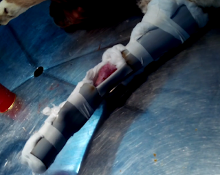
If the kid/ calf cannot stand, splinting the limb in extension is necessary to allow the body weight to load and stretch the tendons and palmar soft tissues. Splints should be applied carefully as they can easily cause pressure sores due to the fragility of the integument and the pressure that is sometimes Necessary to extend the limb. Any strong, light material is suitable for a splint, however, polyvinylchloride (PVC) pipe is readily available and easily cut and shaped to the required size. For neonates, 4-inch diameter thick-walled pipe, cut longitudinally into thirds or halves, is appropriate. The ends of the splint are padded with roll cotton covered with elastic tape. When applying a splint, the limb is bandaged amply, and the splint is positioned over this bandage on the palmar or plantar aspect of the limb. The splints are left on for a maximum of eight h. If re-application of the splints is necessary, the limb should be unsplinted for several hours before the splints are applied again.
A straight splint should be used if the deformity is severe. When the deformity improves, a bend can be placed in the splint at the level of the fetlock joint to further extend this joint. PVC splints can be bent easily by notching each side of the splint and heating it over a flame.
Cooling the splint with cold water will hasten hardening of the splint in the bent position. The use of splints should be discontinued when joint angles approach normal and the animal can stand unassisted. At this point, controlled exercise will usually result in complete correction of the deformity
FIBER GLASS CAST APPLICATION
 A fiberglass cast is the plaster cast made from fiberglass material.  Traditionally,  plaster cast has been made of plaster of Paris. A fiberglass cast is a lighter, synthetic alternative plaster of Paris. Fiberglass is also called glass-reinforced plastic[GRP] or glass fiber reinforced plastic [GFRP] is a fiber reinforced polymer made of a plastic matrix reinforced by fine fibers of glass. Fiberglass cast is a lightweight and  extremely strong material.  As compared to traditional plaster of Paris cast, it is light in weight and  more durable. It is three times stronger and but is only one third in weight. Major orthopaedic ailments encountered in field condition can be effectively addressed using External Coaptation techniques of which FIBER GLASS CASTS can be regarded as the best availiable external coaptation aid.

USE OF ADJUNCT THERAPY 
Correction of the flexural deformity may take several days due to restriction of extension by joint capsule, ligaments, and periarticular tissues. Pain may be marked and analgesics should be administered in the postoperative period. Nonsteroidal anti-inflammatory drugs should be used in the treatment of flexural deformities because the limbs are often painful from stretching the palmar soft tissues. These drugs may cause ulcers, although ketoprofen may be less ulcerogenic than either phenylbutazone or flunixin meglumine. Reducing limb pain and promoting exercise will aid recovery. Balanced nutrition should be ensured for all affected animals. Energy intake should be reduced when much higher than minimal requirements, particularly in rapidly growing kids/ calves

I am sharing some success stories in the form of videos
All these surgeries can be performed in field condition. The main purpose of sharing these videos are to reach the care and attention of healing hands to more number of needy crippled animals, relieve their sufferings and bring back their production life and in turn to make a difference in the life of marginal and small scale farmers.

SURGICAL MANAGEMENT OF BILATERAL FETLOCK KNUCKLING IN CALF 
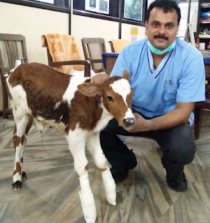
This case was that of a calf presented with bilateral fetlock knuckling . Flexor tenotmies of SFDT was performed on both limbs on the same day of presentation followed by POP bandage. Animal recovered in one week with complete weight bearing. you can watch this video below 


SURGICAL MANAGEMENT OF CARPAL KNUCKLING IN CALF
Carpal knuckling is a difficult one to correct in field condition. generally calves affected with carpal joints are culled with ho hope by the farmer and the vets often don't try to attempt the condition owing to the complicated carpal joint. This case was reffered from Palakkad district and the condition was corrected by a superficial digital flexor tenotomy , ulnaris lateralis partial section and flexor carpi ulnaris tenotomy . Supportive bandage was applied on palmar aspect using modified palmar PVC casts .You can watch this challenging case belpw. 


BILATERAL CARPAL KNUCKLING IN A GOAT KID 
Flexural limb deformities in young calves and kids and Joint Ill are a common orthopedic problem of calves and kids . Deformities of the metacarpophalangeal and distal interphalangeal joints can be mild to severe and no single treatment regime is always successful. A systematic  approach considering the general health ,presenting staus of animal and a treatment plan with deliberate follow ups gives satisfactory results in managing musculoskeletal disorders in calves and kids. 
Bilateral carpal knuckling is a difficult condition to treat in field owing to the high degree of flexion of carpal joints resulting in complete contracture of the carpal joints. The tendon of SFDT and Ulnaris lateralis and Flexor carpi ulnaris needs to be severed to save the animal. This case was a challenging one in which the first surgery was performed on the day of presentation on left fore limb follwed by that the second surgery on day 10 and the complete correction of the carpal knuckling and weight bearing achieved by fourth week of presentation.


Miracles happen every day if we allow it . With a wholehearted dedicated effort, Sincere trials never fail !
Best wishes to all readers of my blog !

Sunday 17 January 2016

GASTRO INTESTINAL FOREIGN BODIES IN DOGS


The gastrointestinal tract consists primarily of the stomach, small intestine and large intestine. It is always difficult to see dogs suffering from foreign body ingestion. But as a Surgeon much excitements and rewarding results are seen in immediate intervention of foreign body ingestion cases . The foreign body entered in side the mouth can pass smoothly through the oesophagus , stomach , small intestines- duodenum, ileum, jejunum, Large intestines- cecum colon rectum and finally it can come through the anal opening outside . But it can also get struck up in any place and the commonest blocks can be encountered in
1.      Pharynx – Isthmus faucum
2.      Oesophagus
3.      Stomach
4.      Duodenum
5.      Jejeunum
6.      Ileum
7.      Ileo cecal junction
In this blog I am sharing some memorable cases of intestinal obstruction in my career.
Due  to indiscriminate eating habits from PICA or Phosphorous deficiency, high worm load due to improper deworming or accidental ingestion of toys or plastic or hard objects , the animal may swallow hard foreign objects which can lodge at any of the above mentioned sites 


NON LINEAR OR SOLID  FOREIGN BODY 
Solid foreign bodies may get lodged in any part of the GIT depends up on the sharpness of edges , size and consistency. There were very two very interesting cases of non linear foreign bodies last month.
TOY WHEEL FOREIGN BODY IN THE JEJUNUM OF A  LABRADOR RETRIEVER 

 A Labrador was referred by a field veterinarian in Thrissur with the history of off feed, not passing motion and extremely painful abdomen. On palpation a hard mass was palpable on the cranial abdomen caudal to the stomach .close examination of  lateral abdomen radiograph revealed a  shadow of  round mass won the abdomen caudal to stomach . On exploratory laparotomy a toy wheel was retrieved from jejunum.  


GRANITE STONE IMPACTION IN A BASSET HOUND

A six year old male Basset hound was presented with symptoms suggestive of chronic pica from the past one month of presentation . The animal was cachectic , anorectic  and was showing recurrent vomiting signs non responsive to medical treatment. On examination a hard mass was palpable on the ventral abdomen and plain radiography revealed a radio opaque mass on the cranial abdomen obliterating the entire lumen and gas filled loops caudal to the obstruction. The animal was stabilised with supplementary fluid therapy
. An exploratory laparotomy was performed by induction and maintenance using thiopentone sodium 2.5 % to the effect under general anaesthesia with atropine and Xylazine as pre medicants @ 0.045 mg /kg bw and and 1 mg / kg bwt respectively.  A hard intestinal segment was isolated at the mid jejunum and a 2 cm long enterotomy incision on non-mesenteric border exteriorised a granite stone piece was exteriorised. Enterotomy closure was carried out using  No 2-0 polyglactic acid suture material in close interrupted pattern. Linea alba was apposed using No 1 poly glactic acid 910 in simple interrupted pattern followed by skin closure in routine manner . Post operatively the animal was treated with antibiotic therapy and fluid therapy for 5 days and oral feeding was withheld for 72 hours post surgery. Sutures were removed on 11th post-operative day and the animal had uneventful recovery.


LINEAR FOREIGNBODY
Gastrointestinal foreign bodies are regarded as main GI emergencies in companion animal practice .Linear foreign bodies, especially strings, can often lead to extreme painful condition and can result in intussusceptions and volvulus which demands immediate surgical intervention.

STORY OF DISCO
I had a dog named Disco when I was working at Hassan Veterinary College. Disco use to accompany me where ever I go and once he came running to me with a long thread seen protruding from his anal opening. On examination a large dressing gauze that he swallowed from the clinics was detected coming out of anus.. One of the student with a good intention to relieve his struggle pulled the thread and he was  crying out of pain .
An immediate laparotomy was performed and a large knot of gauze leading to intussusception (invagination of one part of intestine – intusussipiens to the other part – intususceptum was identified) . The intussusception was corrected and we saved Disco by a timely surgery  

YOU CAN WATCH A VIDEO ON GASTRO INTESTINAL FOREIGN BODIES BELOW 


SIGNS OF OBSTRUCTION
1.    Ingested foreign bodies are more common in young animals                                        
2.    Vomiting is usually seen with an obstructive foreign body of the gastrointestinal tract, however this is a problem that may be seen with many other diseases                                     
3.    Generally if an obstruction of the GI tract is present, diarrhea is usually not a common symptom (as the intestines are obstructed)                                                                             
a.       Loss of appetite                                                                                                                      
b.      Dehydration                                                                                                                               
c.       Abdominal pain                                                                                                               Weight loss may be present if the foreign body is chronic
DIAGNOSIS
1.    Haematology  work is done to determine if there are liver, kidney, pancreas, or electrolyte abnormalities. Complete blood cell count is used to look for signs of infection and anemia.
2.    Radiographs (x-rays) can be very helpful to indicate the presence of a problem in the gastrointestinal tract. Unfortunately, plain radiographs frequently are only suggestive of a problem and do not give us a definitive answer; in some cases the radiographs will be made again to see if the signs of obstruction are repeatable. As a result additional tests such as ultrasound or contrast radiographs (barium swallow) may be indicated.
3.    Sometimes these tests also do not give us a final answer and exploratory surgery is needed.              
4.    If we do not find any obvious problem with the internal organs, biopsies still are done as microscopic disease may be causing the clinical signs.

PRINCIPLES OF INTESTINAL SURGERY
1. Early diagnosis and good surgical technique prevent most complications.
2. Perform surgery as soon as anesthesia is possible in patients with perforation, strangulation, or complete obstruction.
3. Optimal healing requires a good blood supply, accurate mucosal apposition, and minimal surgical trauma.
4. Systemic factors may delay healing and increase the risk of dehiscence, hypovolemia, shock, hypoproteinemia, debilitation, and infection.
5. Use approximating suture patterns: simple interrupted, Gambee, crushing, or simple continuous; stapling techniques are feasible.
6. Engage submucosa in all sutures or staples.
7. Select a monofilament, synthetic absorbable suture such as polydioxanone, polyglyconate,
8. Cover surgical sites with omentum or a serosal patch.
9. Replace contaminated instruments and gloves before closing the abdomen.
10. Fluid Therapy
11. Antibiotic Prophylaxis
12. Assessment of Intestinal Viability
13. Choice of suture material for closure.
14. Monofilament synthetic absorbable (PDS) or synthetic non-absorbable (prolene) are excellent choices.
15. Choice of Suture Pattern
16. Simple interrupted pattern is ideal.
17. Suture Reinforcement
18. Application of Omental and serosal patch aids in faster healing
19. Obtain minimum database: complete blood cell count, chemistry profile, urinalysis, coagulation profile (if possible) with or without an electrocardiogram.
20. Localize the lesion with abdominal palpation radiographs, ultrasonography, and/or endoscopy. Correct hydration, electrolyte, and acid-base abnormalities. Transfuse if the packed cell volume is less than 20% or if the animal is clinically weak or debilitated .Withhold food from mature animals for 12 to 18 hours and from pediatric patients 4 to 8 hours before induction.


 THANKS FOR VISITING MY BLOG 

Friday 23 October 2015

CANINE MAMMARY TUMOR

This segment is intended to give an insight on canine mammary tumors its  direct correlation of the intact uterus and ovaries and the  effect of early  neutering in dogs .

In our university hospital on an average we receive 5-8 cases per week presented with advance canine mammary tumors  . It is a great concern for the dog owners as there is a large awareness among the cancer in human beings. But it is a sad reality that even the most educated pet owner is not fully aware of the direct correlation between the mammary tumor and the intact uterus and ovary in dogs . 

CANINE MAMMARY GLAND - ESSENTIAL  ANATOMY
The number of teats in the dog varies from 8 to 12, with 4 to 6 gland complexes on each side of the midline. Ten is the most common number in larger breeds, four pairs are more common in the smaller breeds. In bitches with ten normal teats, the pattern is two pairs thoracic teats, two pairs abdominal teats, and one pair of inguinal teats.


The number of ducts opening on a teat varies from 8 to 20 external openings per teat for the dog and 1 to 7 for the cat. The openings are located on the blunt end of the teat in an irregular pattern.

The parenchyma, or secretory tissue, is present only during pregnancy, pseudopregnancy, during lactation, and for 40 to 50 days after weaning.
The blood supply of the mammary glands of dogs and cats are similar except for the thoracic glands. In the dog the first pair of thoracic mammary glands receives blood from two sternal branches of the internal thoracic artery, passing between the first and second ribs. The second pair of thoracic mammary glands is supplied by small branches of the mediastinal, or internal mammary, arteries before they anastamose with the sternal branches serving the first pair of glands.

MAMMARY TUMORS
Tumors are frequently seen in the mammary gland of the dog. They may belong to the connective tissue or the epithelial series of mammary tumors, or both. Tumors of the epithelial series are of great importance. Those observed are adenomas, carcinomas, and above all, mixed mammary tumors.
The risk of breast cancer is almost eliminated in dogs that are spayed before their first heat.
Spaying greatly reduces the chances of a female dog developing this condition.
 In those females spayed prior to their first heat cycle, breast cancer is very, very rare. The risk of malignant mammary tumors in dogs spayed prior to their first heat is 0.05%. It is 8% for dog spayed after one heat, and 26% in dogs spayed after their second heat. It is believed that the elimination or reduction of certain hormonal factors causes the lowering of incidence of the disease in dogs that have been spayed. These factors would probably be estrogen, progesterone, a similar hormone or possibly a combination of two or more of these.

The development of mammary gland neoplasms appears to be hormone-dependent because the risk of developing a mammary tumor increases as the number of estrous (heat) cycles increases.

Normal mammary tissue and a majority of benign tumors express both estrogen and progesterone receptors. Less than 50% of mammary carcinomas express either of these receptors. This observation suggests that there is a loss of hormone dependency during transition to malignancy.

Common Signs
Abnormal swellings that persist or continue to grow
Sores that do not heal
Weight loss
Loss of appetite
Bleeding or discharge from any body opening
Offensive odour
Difficulty eating or swallowing
Hesitation to exercise or loss of stamina
Persistent lameness or stiffness
Difficulty breathing, urinating, or defecating

Mammary carcinomas may exhibit rapid growth, doubling in size within a few weeks. However, the size and appearance of these neoplasms can vary greatly. Inflammatory carcinomas usually have diffuse involvement of multiple mammary glands. Edema, erythema, and firmness may be present and affected mammary glands may feel warm to the touch. Dogs with inflammatory carcinoma are more likely to have generalized weakness with anorexia and weight loss. Inflammatory carcinoma is often misdiagnosed as acute mastitis.

TYPE OF TUMORS 
The following are common classes of mammary tumors that might be found on a biopsy.
Fibroadenoma:
A benign glandular tumor for which no treatment is necessary.
Mixed Mammary Tumor:
What is mixed is the type of cell that makes up the tumor: the epithelial cells that line the glandular tissue and the mesenchymal cells that make up the non-glandular portion. (Mixed does not refer to a mix of benign and malignant cells.) The mixed tumor can be either benign or malignant and the biopsy will indicate this.
Adenocarcinoma: 
Adenocarcinomas can be tubular or papillary, depending on the gland cells the tumor arises from. Adenocarcinomas behave malignantly but how aggressively malignant they are depends not on whether they are tubular or papillary, but on other cellular characteristics described by the pathologist (such as how quickly the cells appear to be dividing and how closely they resemble normal gland cells). When the oncologist reads the description he or she will be able to determine how aggressively to combat the tumor.
Inflammatory Carcinoma:
A highly malignant tumor that generates tremendous inflammation locally with ulceration, pus, and discomfort. This type of tumor tends to spread early in its course and is difficult to treat. Fortunately, this especially tragic tumor type accounts for less than 5% of mammary tumors.
In general: approximately 50% of malignant mammary tumors will have already spread by the time of surgery.
The malignant mammary tumors thus in general includes
Tubular adenocarcinomas
Papillary adenocarcinomas
Papillary cystic adenocarcinomas
Solid carcinomas
Anaplastic carcinomas
Osteosarcomas
Fibrosarcomas
Malignant mixed tumors.

Diagnosis of Mammary tumors
Mammary gland tumors are difficult to diagnose by routine cytology and the malignant potential of mammary neoplasms cannot be easily detected in early stages by cytologically.

The cytologic criteria for malignancy in Mammry tumors are

Anisocytosis (variable nuclear size)
Nuclear giant forms
Nuclear or cytoplasmic membrane distortions
High nuclear to cytoplasmic (N:C) ratio
Irregular chromatin shape
Variable chromatin size
Presence of macronucleoli
Variation in nucleolar number
Variation in nucleolar shape
Parachromtin clearing

Hormones 
Approximately 50% of malignant mammary tumors in the dog have receptors for either estrogen or progesterone. This means that the presence of these female hormones promotes the growth of these tumors. Benign tumors also have female hormone receptors and can also be stimulated by hormonal cycling of the female dog. This means that spaying is important even if a tumor has already developed.

MANAGEMENT OF CANINE MAMMARY TUMOR 
Surgical removal of the tumor(s) is the treatment of choice for benign mammary tumors and for malignant mammary tumors that have not spread beyond the mammary tissue and adjacent lymph nodes. If  surgery is done early in the course of this disease, the cancer can be totally eliminated in over 50% of the cases having a malignant form of cancer.Most benign mammary tumors are curable by surgery. Approximately half of malignant mammary tumors are also cured by surgery. This is possible because some malignant mammary tumors in the dog do not spread very quickly and can be removed before they spread. Radical mastectomy has not been shown to be any more effective than more limited surgery. Tumors that are larger than 3 centimeters and tumors that are of higher grade (as classified by the pathologist) are more likely to recur (70% recurrence at 1 year) than smaller tumors and tumors of a lower histopathologic grade (30% recurrence at 1 year). If the dog is not spayed at a young age, spaying at the time of mass removal has recently shown to increase the survival time for some dogs with malignant tumors.
Different terminologies are related to the surgical resection of mammary gland

Mammectomy
Removal of one entire mammary gland, indicated for lesions involving 1/3 of the gland or that are fixed to skin or fascia. Remove skin, abdominal wall fascia.

Regional mastectomy
Removal of affected gland and adjacent mammary glands based on known venous/lymphatic drainage of mammary tissue (may involve axillary, superficial inguinal, sublumbar and cranial sternal nodes).

En bloc mastectomy
Resection of affected glands with the regional lymph node(s)

Unilateral or bilateral (radical) mastectomy
Indicated if multiple tumors or several large tumors along one or both mammary chains, or for any malignant mammary gland tumor in cats.


Chemotherapy: An effective chemotherapy protocol for canine mammary cancer has not been defined. A small percentage of dogs have had remission with drugs such as doxorubicin (Adriamycin) or cisplatin. Because surgery alone is successful in many cases, chemotherapy is usually reserved for tumors that cannot be removed surgically, tumors that have metastasized, and tumors that have a high probability of spreading. Radiation therapy has not been extensively studied to in dogs with mammary tumors. The chemotherapeutic protocol which  can be tried will be a combination of 5-fluorouracil (150 mg/m2 of body surface  area) and cyclophosphamide (100  mg/m2)  but the sideffects masks the results. Postoperative Adjuvant Treatment of Invasive Malignant Mammary Gland Tumors in Dogs with Doxorubicin and Docetaxel has also given excellent results for many practitioners .
A word of caution is always advisable using doxyrubicin . In one case I had encountered a very adverse reaction and tachy cardia associated with doxyrubicin.
Metastasis is a common feature of malignant mammary tumors and chest radiographs will reveal the metastatic nodules in lungs in most of the advanced carcinomas. 
Some cases give real challenges with multiple lesions 
PLEASE WATCH THE VIDEO BELOW 


PREVENTING MAMMARY TUMORS


There is a direct correlation between the early spaying of female dogs and the reduction in the incidence in mammary cancer.
Dogs spayed before coming into their first heat have an extremely small chance of ever developing mammary cancer. 
Dogs spayed after their first heat but before 2.5 years are at more risk, but less risk than that of dogs who were never spayed, or spayed later in life. As a veterinarian I strongly believe that .......MAMMARY TUMORS ARE  PREVENTABLE  BY EARLY SPAYING .