This segment
is intended to give an insight on canine mammary tumors its direct correlation of the intact uterus and
ovaries and the effect of early neutering in dogs .

 Normal
mammary tissue and a majority of benign tumors express both estrogen and
progesterone receptors. Less than 50% of mammary carcinomas express either of
these receptors. This observation suggests that there is a loss of hormone
dependency during transition to malignancy.
Normal
mammary tissue and a majority of benign tumors express both estrogen and
progesterone receptors. Less than 50% of mammary carcinomas express either of
these receptors. This observation suggests that there is a loss of hormone
dependency during transition to malignancy.
 Abnormal
swellings that persist or continue to grow
Abnormal
swellings that persist or continue to grow
 Adenocarcinoma:
Adenocarcinomas can be tubular or papillary, depending on the gland cells the tumor arises from. Adenocarcinomas behave malignantly but how aggressively malignant they are depends not on whether they are tubular or papillary, but on other cellular characteristics described by the pathologist (such as how quickly the cells appear to be dividing and how closely they resemble normal gland cells). When the oncologist reads the description he or she will be able to determine how aggressively to combat the tumor.
 Surgical removal of the tumor(s) is the treatment
of choice for benign mammary tumors and for malignant mammary tumors that have
not spread beyond the mammary tissue and adjacent lymph nodes. If surgery is done early in the course of this
disease, the cancer can be totally eliminated in over 50% of the cases having a
malignant form of cancer.Most benign mammary tumors are curable by surgery. Approximately half of
malignant mammary tumors are also cured by surgery. This is possible because
some malignant mammary tumors in the dog do not spread very quickly and can be
removed before they spread. Radical mastectomy has not been shown to be any
more effective than more limited surgery. Tumors that are larger than 3 centimeters
and tumors that are of higher grade (as classified by the pathologist) are more
likely to recur (70% recurrence at 1 year) than smaller tumors and tumors of a
lower histopathologic grade (30% recurrence at 1 year). If the dog is not
spayed at a young age, spaying at the time of mass removal has recently shown
to increase the survival time for some dogs with malignant tumors.
Surgical removal of the tumor(s) is the treatment
of choice for benign mammary tumors and for malignant mammary tumors that have
not spread beyond the mammary tissue and adjacent lymph nodes. If surgery is done early in the course of this
disease, the cancer can be totally eliminated in over 50% of the cases having a
malignant form of cancer.Most benign mammary tumors are curable by surgery. Approximately half of
malignant mammary tumors are also cured by surgery. This is possible because
some malignant mammary tumors in the dog do not spread very quickly and can be
removed before they spread. Radical mastectomy has not been shown to be any
more effective than more limited surgery. Tumors that are larger than 3 centimeters
and tumors that are of higher grade (as classified by the pathologist) are more
likely to recur (70% recurrence at 1 year) than smaller tumors and tumors of a
lower histopathologic grade (30% recurrence at 1 year). If the dog is not
spayed at a young age, spaying at the time of mass removal has recently shown
to increase the survival time for some dogs with malignant tumors.
 Chemotherapy:
An effective chemotherapy protocol for canine mammary cancer has not been
defined. A small percentage of dogs have had remission with drugs such as
doxorubicin (Adriamycin) or cisplatin. Because surgery alone is successful in
many cases, chemotherapy is usually reserved for tumors that cannot be removed
surgically, tumors that have metastasized, and tumors that have a high
probability of spreading. Radiation therapy has not been extensively studied to
in dogs with mammary tumors. The chemotherapeutic protocol which can be tried will be a combination of 5-fluorouracil
(150 mg/m2 of body surface area) and
cyclophosphamide (100 mg/m2) but the sideffects masks the results. Postoperative
Adjuvant Treatment of Invasive Malignant Mammary Gland Tumors in Dogs with
Doxorubicin and Docetaxel has also given excellent results for many
practitioners .
Chemotherapy:
An effective chemotherapy protocol for canine mammary cancer has not been
defined. A small percentage of dogs have had remission with drugs such as
doxorubicin (Adriamycin) or cisplatin. Because surgery alone is successful in
many cases, chemotherapy is usually reserved for tumors that cannot be removed
surgically, tumors that have metastasized, and tumors that have a high
probability of spreading. Radiation therapy has not been extensively studied to
in dogs with mammary tumors. The chemotherapeutic protocol which can be tried will be a combination of 5-fluorouracil
(150 mg/m2 of body surface area) and
cyclophosphamide (100 mg/m2) but the sideffects masks the results. Postoperative
Adjuvant Treatment of Invasive Malignant Mammary Gland Tumors in Dogs with
Doxorubicin and Docetaxel has also given excellent results for many
practitioners .
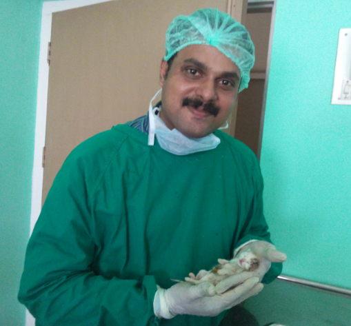
In our
university hospital on an average we receive 5-8 cases per week presented with
advance canine mammary tumors . It is a
great concern for the dog owners as there is a large awareness among the cancer
in human beings. But it is a sad reality that even the most educated pet owner
is not fully aware of the direct correlation between the mammary tumor and the
intact uterus and ovary in dogs .
CANINE MAMMARY
GLAND - ESSENTIAL ANATOMY
The number of
teats in the dog varies from 8 to 12, with 4 to 6 gland complexes on each side
of the midline. Ten is the most common number in larger breeds, four pairs are
more common in the smaller breeds. In bitches with ten normal teats, the
pattern is two pairs thoracic teats, two pairs abdominal teats, and one pair of
inguinal teats.

The number of
ducts opening on a teat varies from 8 to 20 external openings per teat for the
dog and 1 to 7 for the cat. The openings are located on the blunt end of the
teat in an irregular pattern.
The
parenchyma, or secretory tissue, is present only during pregnancy,
pseudopregnancy, during lactation, and for 40 to 50 days after weaning.
The blood
supply of the mammary glands of dogs and cats are similar except for the
thoracic glands. In the dog the first pair of thoracic mammary glands receives
blood from two sternal branches of the internal thoracic artery, passing
between the first and second ribs. The second pair of thoracic mammary glands
is supplied by small branches of the mediastinal, or internal mammary, arteries
before they anastamose with the sternal branches serving the first pair of
glands.
MAMMARY
TUMORS
Tumors are
frequently seen in the mammary gland of the dog. They may belong to the
connective tissue or the epithelial series of mammary tumors, or both. Tumors
of the epithelial series are of great importance. Those observed are adenomas,
carcinomas, and above all, mixed mammary tumors.
The risk of breast cancer is almost eliminated in
dogs that are spayed before their first heat.
Spaying greatly reduces the chances of a female dog developing this condition.
Spaying greatly reduces the chances of a female dog developing this condition.
In
those females spayed prior to their first heat cycle, breast cancer is very,
very rare. The risk of malignant mammary tumors in dogs spayed prior to their
first heat is 0.05%. It is 8% for dog spayed after one heat, and 26% in
dogs spayed after their second heat. It is believed that the elimination or
reduction of certain hormonal factors causes the lowering of incidence of the
disease in dogs that have been spayed. These
factors would probably be estrogen, progesterone, a similar hormone or possibly
a combination of two or more of these.
The
development of mammary gland neoplasms appears to be hormone-dependent because
the risk of developing a mammary tumor increases as the number of estrous
(heat) cycles increases.
 Normal
mammary tissue and a majority of benign tumors express both estrogen and
progesterone receptors. Less than 50% of mammary carcinomas express either of
these receptors. This observation suggests that there is a loss of hormone
dependency during transition to malignancy.
Normal
mammary tissue and a majority of benign tumors express both estrogen and
progesterone receptors. Less than 50% of mammary carcinomas express either of
these receptors. This observation suggests that there is a loss of hormone
dependency during transition to malignancy.
Common Signs
 Abnormal
swellings that persist or continue to grow
Abnormal
swellings that persist or continue to grow
Sores that do
not heal
Weight loss
Loss of
appetite
Bleeding or
discharge from any body opening
Offensive
odour
Difficulty
eating or swallowing
Hesitation to
exercise or loss of stamina
Persistent
lameness or stiffness
Difficulty
breathing, urinating, or defecating
Mammary
carcinomas may exhibit rapid growth, doubling in size within a few weeks.
However, the size and appearance of these neoplasms can vary greatly.
Inflammatory carcinomas usually have diffuse involvement of multiple mammary
glands. Edema, erythema, and firmness may be present and affected mammary
glands may feel warm to the touch. Dogs with inflammatory carcinoma are more
likely to have generalized weakness with anorexia and weight loss. Inflammatory
carcinoma is often misdiagnosed as acute mastitis.
TYPE OF TUMORS
The following are common classes of mammary tumors that might be found on a biopsy.
The following are common classes of mammary tumors that might be found on a biopsy.
Fibroadenoma:
A benign glandular tumor for which no treatment is necessary.
A benign glandular tumor for which no treatment is necessary.
Mixed Mammary Tumor:
What is mixed is the type of cell that makes up the tumor: the epithelial cells that line the glandular tissue and the mesenchymal cells that make up the non-glandular portion. (Mixed does not refer to a mix of benign and malignant cells.) The mixed tumor can be either benign or malignant and the biopsy will indicate this.
What is mixed is the type of cell that makes up the tumor: the epithelial cells that line the glandular tissue and the mesenchymal cells that make up the non-glandular portion. (Mixed does not refer to a mix of benign and malignant cells.) The mixed tumor can be either benign or malignant and the biopsy will indicate this.
 Adenocarcinoma:
 Adenocarcinomas can be tubular or papillary, depending on the gland cells the tumor arises from. Adenocarcinomas behave malignantly but how aggressively malignant they are depends not on whether they are tubular or papillary, but on other cellular characteristics described by the pathologist (such as how quickly the cells appear to be dividing and how closely they resemble normal gland cells). When the oncologist reads the description he or she will be able to determine how aggressively to combat the tumor.
Inflammatory Carcinoma:
A highly malignant tumor that generates tremendous inflammation locally with ulceration, pus, and discomfort. This type of tumor tends to spread early in its course and is difficult to treat. Fortunately, this especially tragic tumor type accounts for less than 5% of mammary tumors.
A highly malignant tumor that generates tremendous inflammation locally with ulceration, pus, and discomfort. This type of tumor tends to spread early in its course and is difficult to treat. Fortunately, this especially tragic tumor type accounts for less than 5% of mammary tumors.
In general:
approximately 50% of malignant mammary tumors will have already spread by the
time of surgery.
The malignant mammary tumors thus in general includes
The malignant mammary tumors thus in general includes
Tubular
adenocarcinomas
Papillary
adenocarcinomas
Papillary
cystic adenocarcinomas
Solid
carcinomas
Anaplastic
carcinomas
Osteosarcomas
Fibrosarcomas
Malignant
mixed tumors.
Diagnosis of Mammary
tumors
Mammary gland
tumors are difficult to diagnose by routine cytology and the malignant
potential of mammary neoplasms cannot be easily detected in early stages by cytologically.
The cytologic
criteria for malignancy in Mammry tumors are
Anisocytosis
(variable nuclear size)
Nuclear giant
forms
Nuclear or
cytoplasmic membrane distortions
High nuclear to
cytoplasmic (N:C) ratio
Irregular
chromatin shape
Variable
chromatin size
Presence of
macronucleoli
Variation in
nucleolar number
Variation in
nucleolar shape
Parachromtin
clearing
Hormones
Approximately
50% of malignant mammary tumors in the dog have receptors for either estrogen
or progesterone. This means that the presence of these female hormones promotes
the growth of these tumors. Benign tumors also have female hormone receptors
and can also be stimulated by hormonal cycling of the female dog. This means
that spaying is important even if a tumor has already developed.
MANAGEMENT OF CANINE MAMMARY TUMOR
 Surgical removal of the tumor(s) is the treatment
of choice for benign mammary tumors and for malignant mammary tumors that have
not spread beyond the mammary tissue and adjacent lymph nodes. If surgery is done early in the course of this
disease, the cancer can be totally eliminated in over 50% of the cases having a
malignant form of cancer.Most benign mammary tumors are curable by surgery. Approximately half of
malignant mammary tumors are also cured by surgery. This is possible because
some malignant mammary tumors in the dog do not spread very quickly and can be
removed before they spread. Radical mastectomy has not been shown to be any
more effective than more limited surgery. Tumors that are larger than 3 centimeters
and tumors that are of higher grade (as classified by the pathologist) are more
likely to recur (70% recurrence at 1 year) than smaller tumors and tumors of a
lower histopathologic grade (30% recurrence at 1 year). If the dog is not
spayed at a young age, spaying at the time of mass removal has recently shown
to increase the survival time for some dogs with malignant tumors.
Surgical removal of the tumor(s) is the treatment
of choice for benign mammary tumors and for malignant mammary tumors that have
not spread beyond the mammary tissue and adjacent lymph nodes. If surgery is done early in the course of this
disease, the cancer can be totally eliminated in over 50% of the cases having a
malignant form of cancer.Most benign mammary tumors are curable by surgery. Approximately half of
malignant mammary tumors are also cured by surgery. This is possible because
some malignant mammary tumors in the dog do not spread very quickly and can be
removed before they spread. Radical mastectomy has not been shown to be any
more effective than more limited surgery. Tumors that are larger than 3 centimeters
and tumors that are of higher grade (as classified by the pathologist) are more
likely to recur (70% recurrence at 1 year) than smaller tumors and tumors of a
lower histopathologic grade (30% recurrence at 1 year). If the dog is not
spayed at a young age, spaying at the time of mass removal has recently shown
to increase the survival time for some dogs with malignant tumors.
Different
terminologies are related to the surgical resection of mammary gland
Mammectomy
Removal of
one entire mammary gland, indicated for lesions involving 1/3 of the gland or
that are fixed to skin or fascia. Remove skin, abdominal wall fascia.
Regional mastectomy
Removal of
affected gland and adjacent mammary glands based on known venous/lymphatic
drainage of mammary tissue (may involve axillary, superficial inguinal, sublumbar
and cranial sternal nodes).
En bloc mastectomy
Resection of
affected glands with the regional lymph node(s)
Unilateral or bilateral (radical) mastectomy
Indicated if
multiple tumors or several large tumors along one or both mammary chains, or
for any malignant mammary gland tumor in cats.
 Chemotherapy:
An effective chemotherapy protocol for canine mammary cancer has not been
defined. A small percentage of dogs have had remission with drugs such as
doxorubicin (Adriamycin) or cisplatin. Because surgery alone is successful in
many cases, chemotherapy is usually reserved for tumors that cannot be removed
surgically, tumors that have metastasized, and tumors that have a high
probability of spreading. Radiation therapy has not been extensively studied to
in dogs with mammary tumors. The chemotherapeutic protocol which can be tried will be a combination of 5-fluorouracil
(150 mg/m2 of body surface area) and
cyclophosphamide (100 mg/m2) but the sideffects masks the results. Postoperative
Adjuvant Treatment of Invasive Malignant Mammary Gland Tumors in Dogs with
Doxorubicin and Docetaxel has also given excellent results for many
practitioners .
Chemotherapy:
An effective chemotherapy protocol for canine mammary cancer has not been
defined. A small percentage of dogs have had remission with drugs such as
doxorubicin (Adriamycin) or cisplatin. Because surgery alone is successful in
many cases, chemotherapy is usually reserved for tumors that cannot be removed
surgically, tumors that have metastasized, and tumors that have a high
probability of spreading. Radiation therapy has not been extensively studied to
in dogs with mammary tumors. The chemotherapeutic protocol which can be tried will be a combination of 5-fluorouracil
(150 mg/m2 of body surface area) and
cyclophosphamide (100 mg/m2) but the sideffects masks the results. Postoperative
Adjuvant Treatment of Invasive Malignant Mammary Gland Tumors in Dogs with
Doxorubicin and Docetaxel has also given excellent results for many
practitioners .
A
word of caution is always advisable using doxyrubicin . In one case I had encountered
a very adverse reaction and tachy cardia associated with doxyrubicin.
Metastasis
is a common feature of malignant mammary tumors and chest radiographs will
reveal the metastatic nodules in lungs in most of the advanced carcinomas.
Some cases give real challenges with multiple lesions
PLEASE WATCH THE VIDEO BELOW
PREVENTING MAMMARY TUMORS
There is a direct correlation between the early spaying of
female dogs and the reduction in the incidence in mammary cancer.
Dogs spayed
before coming into their first heat have an extremely small chance of ever
developing mammary cancer.
Dogs spayed after their first heat but before 2.5
years are at more risk, but less risk than that of dogs who were never spayed,
or spayed later in life. As a veterinarian I strongly believe that .......MAMMARY TUMORS ARE PREVENTABLE BY EARLY SPAYING .