I am sharing some insights on the Feline Dystocia and C- Section
in this segment .
Many times we get cats presented
in advanced stage of dystocia. It is very important for the cat owners,
Paravets to understand the stage of birth which clearly indicates the time for
intervention by the vet.
Cat’s labour signs are very
interesting and much more unique compared to canine labour stages .
STAGE -1 First stage labour begins with the onset of uterine contractions.
During this stage the cervix begins to dilate open with clear , odourless
discharge from the vagina - mucus plug during pregnancy sealing the uterus starts.
As the first stage progresses, contractions will become closer and closer
together.
STAGE -2 Second stage labour is characterised
by contractions much stronger and closer together and the cervix becomes fully
dilated. When the queen is ready , kitten moves down the birth canal.
Pressure on the cervix initiates an urge to push from the mother. It
takes around three pushes for the kitten to be delivered. The queen will tear
and lick the membranes from the face and body which will stimulate breathing. Second
stage usually takes around 5 minutes to 1 hour.
MORE THAN ONE HOUR OF SECOND
STAGE OF LABOUR NEED VETERINARY HELP!
STAGE -3 Third stage labour: Immediately
following the kitten's birth, the placenta is normally delivered. Once the
queen has cleaned the kitten and breathing has commenced normally the queen
will chew the umbilical cord in two and quite often will eat the placenta.
CAUSES OF DYSTOCIA IN CAT
There are a number of causes of dystocia which can
be classified as maternal or fetal. Maternal causes include Narrow pelvis. Fractured pelvis ,Pelvic
obstruction and Uterine inertia uterine torsion (twisting) or rupture. This may
be primary or secondary . When the uterus fails to produce any contractions this
is regarded as Primary inertia This can be from poor health, obesity, age, stress.Fetal causes include abnormally
presentation of kitten, oversized foetus
, Excessive fetal head size and malformed fetus, Fetal death.
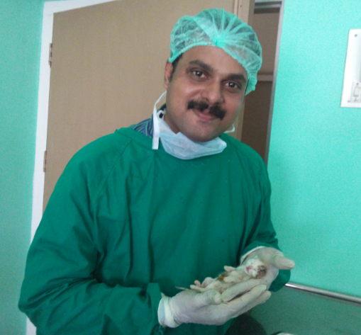
A CASE OF UTERINE TORSION LEADING TO DYSTOCIA AND REVIVAL OF LIVE KITTEN BY CESAREAN SECTION
PLEASE WATCH THE VIDEO BELOW .
We had handled a very challenging case of feline dystocia this week. three year old cat was presented on 71 day of gestation with history of delivering still born kitten 11 hrs prior to presentation of case
.The case was tried to manage medically with oxytocin but the total uterine inertia was evidentThe cat was very dull and depressed. An abdominal ultrasound revealed two more kittens on the right horn. One dead and another alive. An emergency C- Section was planned .
On laprotomy , uterine torsion of gravid right horn and pale purple color of the uterine mass was suggestive of putrefaction . On opening the uterus a very foul smelling foetus was removed and a live kitten was carefully taken.
Upon removal the kitten had gone for apnoea which was successfully retrieved by cardio pulmonary resuscitation .
THE JOY OF SAVING A MOTHER AND A KITTEN WAS BEYOND WORDS TO EXPRESS .
SIGNS OF DYSTOCIA IN CATS
·Gestation lasting longer than 70 days
·Stage 1 labour lasting longer than 24 hours
·Straining for 10 minutes if a fetus is visible in the birth canal
·Acute depression and Fever (above 103°F)
·Sudden discharge from the vagina of bright red blood lasting longer than 10 minutes
·Thick, black, foul smelling discharge from the vagina.
Significant gap between deliveries is very common in cats . If the fetus is stuck in the birth canal, apply gentle traction is needed . An ultrasound scan can precisely diagnose the live kitten in the abdomen . In my experience I had found that If the labour has been long, calcium levels may have depleted. When combined with oxytocin, calcium gluconate can result in contractions much more efficient than with oxytocin alone. Adequate hydration with Intravenous fluids is a must to combat the stress of dystocia.
And when the queen is not responding to medical treatment a Cesarean section is the only resort.
Anesthetic combination of Ketamine @ 15 mg / kg bw and diazepam @ 0.6 mg/kg bw gives very good result during the procedure. Ketamine in a slight higher dose were found to be showing prolonged recovery period and catalepsy. In case of Dystocias delivering one dead foetus and mother still straining .. An ultrasound scan is an excellent tool to identify the live foetus if any and in that case only an emergency C Section can save the life of the mother and kitten.
As
an Army Veterinarian I had spent my majority time with Horses and Mules , I had
an impression that Donkeys are like horses and I can very well treat them when
given a chance. All my convictions were proved wrong when I got the real life challenge
to treat a donkey in advanced state of colic. Thanks to the the wonderful
animal welfare organisations, Animal Rahat- PETA and Donkey Sanctuary International (an
International donkey welfare organization) which taught me much regarding this
unique versatile creation of god. Donkeys are definitely not like horses. In
many ways they have a complex psycho somatic difference and very high level of
pain tolerance , making a vet very difficult to interpret the concealed
symptoms of diseases . During my graduation days much less were taught regarding the care, management and diseases of donkeys. Even today the VCI syllabus addresses very less or nil regarding the
donkeys.
I
would like to dedicate this segment to the vets and animal welfare
professionals who work for donkeys. I believe their work is indeed noblest of
all . God has created donkeys with elements of extra dose of tolerance,
resilience and much versatility.
Donkeys
belongs to the class equidae . Much similar to the domestic cousin- Horse and wild relative
zebra, donkey possess its own individuality and
character making him poor man’s friend and ultimately “ beast of burden”.
Donkeys are agile, firm and much resilient animals with a great deal of
tolerance and complacence. The donkeys differ in horses in following ways . Chromosome
number of a horse is 64, Donkey has 62 chromosomes (Zebra has chromosome number
46).
Donkeys
are not like horses; they differ physically, mentally and emotionally.
More
stoical in their
behaviour , donkeys show
limited fear response
to novel situations
and this can
be mistaken for stubbornness rather than fear.
Donkeys (Equus asinus) are very
efficient at metabolising their food . Donkeys form very
strong bonds with other
donkeys and animals,
and even short term separation from a companion can be
stressful.
Donkey ergots are more prominent than for horses– Often look more like a
digital pad
Donkey inferior check ligaments– Have
an extension from the deep flexor tendon to the superficial flexor tendon in
the front legs (not found in the mule).
No Inf Check Ligament in the rear
leg
Laryngeal anatomy slightly differs
from horses and nasal passages are
smaller than equal sized horses – Smaller NG tubes required
Gestation period of a donkey is 370
were as for horses it is 340. Average life span of donkey is 35 were as for
horses it is 25.
Crossing a donkey with a horse produce
Mules and Hinnies.
Mule- female
horse bred to a male ass- 63 chromosomes
Hinny- male horse bred to a female ass- 63
chromosomes also Both crosses are considered sterile even though there are
documented cases of fertility in the female mule (Mare Mule or Molly Mule).
THE REAL LIFE OF
INDIAN DONKEYS
I made a small clip about the life of donkeys in India from the collection of photographs that I gathered during all these days.
YOU CAN WATCH THE CLIP BELOW
Large number of donkeys are bred and exploited as pack
animals in our country especially in North India. Donkeys are mainly used in
brick klins. In south you can see a great number of Donkeys in Maharashtra-
Sangli and Solapur district and many areas of Tamil Nadu. Much compact body, a
docile nature, a “no problem “,“no complaints” attitude makes a donkey -poor
man’s friend and beast of burden.
When it comes to
tolerance , heaven is the limit for donkeys . It is absolutely true when we say
that India's infrastructure was built off their forced servitude in the brick
industry. Donkeys are routinely overloaded; young, undersized and ill or
injured animals frequently haul loads of more than 50 bricks with little to no
padding between the load and their backs. As a result, donkeys can develop
chafing wounds, bruises and abrasions which can become infected and infested
with maggots. Their hooves can become painfully overgrown, cracked and sore,
and lameness is exceedingly common.
Donkeys can suffer from a host of diseases, including rabies,
trypanosomiasis, extensive dermatitis, ophthalmic infections and a wide variety
of polythene impactive colics (which may not be prevalent outside India). Stoic
and acquiescent, donkeys are easy targets for abuse, and their suffering often
goes unnoticed. Impoverished
people with little knowledge of humane husbandry methods perpetuate many cruel
practices against these animals. Donkeys have their nostrils cut open,
ostensibly to increase air flow, and their ears are also cut open to drain
what's perceived as "bad blood" and to prevent tetanus. Most donkeys
never see a veterinarian during their entire lifetime, and there is little
scientific research pertaining to the health and welfare of donkeys in India. I was shocked to see donkeys been used for drawing sand from river bed . These " water donkeys" end up their life with irreparable hoof decay and wide variety of hoof ailments.
Illegal attempts to transport donkeys across the borders of Maharashtra and
Karnataka to be killed for meat are still reported. As evidenced by the above,
donkeys are among the most abused animals on Earth.
When I was working
for PETA as Program Manager and Chief Vet Surgeon for the Animal Rahat , the
largest working animal welfare program, as a team we learned a lot
treating wide ailments in donkeys.
Understanding the socio economic structure of a donkey owners life is the first
step in understanding the physical condition of donkeys. In majority of brick klins of our country , It is a common
scene to watch 3 year old kids grossly malnourished leading a 2 year old donkey colt
over loaded with bricks. If you go to his hut you can see his ailing carrying mother with five
more hungry kids playing around in a filthy, totally unhygienic environment.
What is the role of animal welfare in the place where human welfare is grossly
compromised ?
Tetanus
and Rabies are the most dangerous disease conditions in donkeys apart from
plastic colics , gross worm infestations and wide range of wounds and hoof
affections. The sight of the donkey with rabies is really horrific to watch and
much challenging even to approach for
euthanasia. An average donkey needs regular deworming with broad-spectrum
dewormers – Fenbendazole and praziquantel combinations , regular hoof cleaning
and body grooming apart from nutritious
diet of plenty of green grass, chaffs , wheat bran and protein sources . But in
real field scenario donkeys graze on road side eating plastics invariably
leading to plastic colics.
Colic in donkeys requires special attention as the
only pain signs in donkey with colic will be anb increase in heart beat and
respiration rate. The tolerance of this species is too high that many clinical
signs get concealed.
Where
ever we see donkey owners ,it is a mandate to advice them for tetanus toxoid
injection and anti rabies injection annually.
A
WAY FOREWARD
I
strongly believe that Animal Welfare is the extension of Human welfare and this
is more pronounced with donkey welfare. As the donkey owners are the poorest of
all animal handlers who live with gross poverty , difficult and struggling to
earn their routine bread . It is a very
common fact that literacy rate is almost
nil with donkey owners. Poverty breeds callous attitude and we can see animal welfare
being compromised grossly in brick klins in the country. If we really want to
make some difference to this condition of donkeys we need to work a lot at the
community level and a great deal of society upliftment is the only way to bring
welfare for this poor creatures. In a country were crores of money being pumped
in the name of CSR, I believe it is the duty of every corporate, every building
built on brick to address this sufferings of this beast of burden. In other way
I would like to put as“ for every brick produced in a brick kiln we owe its
share to the mighty donkeys who worked in the brick klins”. A veterinarian role
in community development is much
pronounced in practice of donkey
medicine.
I thank the wonderful team of Donkey Sanctuary
India and UK, Animal Rahat PETA , Brookes India who laid foundation for this
great initiatives for struggling donkeys in our country. The community
development programmes by these organisations had literally made paradigm shift
in the approach towards the donkeys .
I salute all my fellow veterinarians and
animal welfare staff in these organizations and in field who works hard to
address the suffering of the donkeys. I am equally proud and honored to be one
amongst you who care a lot for this wonderful creation of god .
I believe a lot in miracles. Nothing ever happen by chance and there is a time and reason for every single leaf falling on ground. Many times a single action of kindness bring back the most hopeless condition in an animal's life. I strongly believe that all we need are eyes and heart which are " truly alive". The joy of bringing back a wagging tail to life is beyond words to express.
This
is another rescue story of a puppy.
This homeless pup was found abandoned, crying with pain on road side with a huge painful swelling on the inguinal
region. We rescued this pup and an emergency herniorrhaphy under a well planned
balanced anesthetic regime saved the life of the pup. The puppy had an uneventful recovery . Inguinal hernia needs immediate attention. In pups this starts as a swelling on the groin and majority cases this will be congenital. As age advances the prognosis of the condition worsens. This puppy was identified with a congenital inguinal hernia. He responded well to the surgery.
You
can watch this video below
The
two important abdominal hernias in puppies are Umbilical hernia and Inguinal
hernia. An inguinal hernia is a condition in which the
abdominal contents protrude through the inguinal canal or
inguinal ring, an opening which occurs in the muscle wall in the groin area.
The muscles
Sartorius ,Pectineus , Adductor and fascia of external abdominis obliques forms
the external inguinal ring boundary. In congenital development defect, the
inguinal ring will be ill developed making the intestine and viscera herniating
through the improperly closed external
inguinal ring.
As the puppy start growing the hernia
contents increases and strangulation at the level of the ring is a common
sequelae. The condition is severely painful. Early identification of case and
herniorrhaphy is the only treatment option.
Symptoms
seen with a complicated inguinal hernia may include: Swelling in the groin
area, which may become painful and warm to the touch, pain, frequent attempts
to urinate, lack of appetite and depression, the situation can become
life-threatening.
Inguinal
hernias can usually be diagnosed by finding the swelling caused by the hernia
on a physical examination. However, sometimes contrast radiographs (X-rays) or
an abdominal ultrasound are needed to determine which abdominal contents, if
any, are entrapped.
Herniorrhaphy
is the only solution and the success of the surgery is more in young animals.
Interrupted sutures with chromic catgut or polyglactin 910 suture materials follwoed by a subcuticular sutures give sufficient tension handling of the inguinal ring. Balanced anesthetic protocol with premedication with triflupromazine followed by induction with a higher dilution of 12.5 % thio pentone soduim and maintenance with isoflurane or sevoflurane anesthesia is the best to deal with inguinal hernia correction in puppies.
The joy of recovery is beyond words to express. The puppy got attached with me and all those budding vets ., the internees who took care of him very well till recovery. I feel blessed and whole heartedly thank god for making me capable to save innocent lives.
The stain of pain and fear is rooted in all living beings . A little gesture and a thoughtful moment by our actions and deeds brings unbelievable difference in the lives of the animals. I am certain that every one who read this segment will definitely agree with me and spread the small yet invaluable message of kindness and compassion.
Until he extends the circle of his compassion to all living things, man will not himself find peace. -Albert Schweitzer
In this
post, I am sharing some blessed moments of my career as a vet surgeon. Last week a case of 4 yr old
biparous Labrador retriever in 64th day of gestation was presented in
total refractory state of complete uterine inertia with history of straining
from past 24 hours . The owner brought this dog in a very late stage and the
animal had severe dehydration, cachexia and stage of unconsciousness. After initial
stabilization with fluid therapy , an emergency cesarean was performed which saved the animal and nine live pups were successfully
revived
Even
though this operation was done with out assessing the foetal position and
number by an ultrasonic scan due to the
late night presentation of the case , the animal responded very well and the
anesthetic ombination of propofol @ 6 mg/kg
and diazepam @ 0.5 mg/kg gave excellent results.
PLEASE WATCH THE VIDEO BELOW
Dystocia
can occur as a consequence of problems with the dam’s uterus or birth canal, or
with the fetus. An abnormally long time
between deliveries of puppies, prolonged
period of labor with no puppies being born, delivery of stillborn puppies (born
dead),presence of a puppy in the birth canal without obvious abdominal
contractions or movement of the puppy, agitation in the dam, persistent nesting
behavior (digging, circling),fatigue, exhaustion, Straining as if to defecate ,
suggests an obstruction in the birth canal or uterine inertia which demands a
Cesarean section to save the mother as well as the pups.
The
diagnosis of dystocia should be based on the presence of any of the following
criteria:
Failure
of the dam to initiate labor at term.
Bitches can be considered over term at >70-72 days from the first
breeding, >58-60 days of diestrus, or > 66 days from the LH surge or
initial rise in progesterone during estrus (All ovulation timing methods
performed during the previous cycle).
Failure
of the dam to enter Stage 1 labor, beyond 24-36 hours after a detectable drop
in rectal temperature <99-100 degrees F or decline in progesterone level
below 2 ng/ml, or failure to proceed from Stage 1 to Stage 2 labor within 24
hours.
Failure
of the dam to complete delivery of all of the fetuses in a timely fashion. Delivery of the first fetus should occur
within 1 hour of active labor (with visible abdominal efforts) or 4-6 hours of
intermittent labor.
Fetal
distress (unborn puppies with slow heart rates, stillbirths).
Maternal
distress (excessive pain or depression), green or copious vaginal bleeding.
Irreversible
history of dystocia (pelvic canal abnormalities, mismatch fetal/maternal size, malposition)
In
the present case the mother was totally non responsive and cachectic with
Severe
straining. The owner did gross neglect to the mother leading to the
presentation of case in advance stage.
Oxytocin
injection can be given if the mother is in a position to initiate a contraction.
In this case emergency fluid therapy stabilized the animal.
My previous post on C-Section in a PUG can also be refereed in this context.
Patients
who present for dystocia often will be dehydrated, so it is appropriate to
fluid-resuscitate them before surgery. Electrolyte abnormalities should be
corrected pre-operatively. Every animal must be evaluated for evidence of
hypovolemic shock, and treated appropriately if necessary. The vast majority of
animals, however, will require only one-third to one-quarter of their shock
dose of isotonic crystalloid fluid (15-20 ml/kg), followed by a surgical rate
of fluid administration (10 ml/kg/hr crystalloid unless complicating factors
are present, such as heart disease or hypoproteinemia).
Nearly
all analgesic and anesthetic drugs will cross the placenta and enter the fetal
circulation. As such, it is important to minimize fetal exposure to
cardiovascular depressant drugs by minimizing time from induction to delivery.
PROPOFOL
-DIAZEPAM COMBINATIONS FOR ANESTHETIC MANAGEMENT IN C- SECTION.
Propofol
is an anesthetic injection for use in dogs for induction and maintenance of
general anesthesia for up to 20 minutes and for induction where maintenance is
provided by inhalant anesthetics.
When using propofol, patients should be continuously monitored, and facilities
for the maintenance of a patent airway, artificial ventilation and oxygen
supplementation must be immediately available. Propofol contains no
antimicrobial preservatives. Strict aseptic techniques must always be
maintained during handling since the vehicle is capable of supporting the rapid
growth of microorganisms.
Propofol
works by binding to the GABA receptor (the primary inhibitory neurotransmitter
binding site) CNS Effects Propofol produces CNS depression and unconsciousness.
Propofol is considered “cerebral-friendly” because it decreases cerebral oxygen
consumption, decreases cerebral blood flow (CBF), lowers intra-cranial pressure
.Administration of other GABA agonists (e.g. benzodiazepines (diazepam/
midazolam)) immediately before propofol can decrease the amount of propofol
needed by almost 50%. This is cost saving, and also decreases the
cardiovascular and respiratory depression seen with bigger doses of propofol.
I
thank my colleague Dr Nagaraj, the internees in Veterinary Surgery and the final B.V.Sc. students who helped me in
this procedure.Hope this case has inspired the budding vets . I am sure my team will remember this case through out the career
for the timely decision to cut open and the joy of taking live pups one by one and counting the heartbeats reviving nine pups sequentially was beyond the words to express.
THANKS FOR VISITING MY BLOG
You can subscribe my youtube channel for more clinical veterinary surgery videos
In this segment I would like to share some experience with Canine testicular tumors- sertoli cel tumor and seminoma which is a testosterone dependent tumor seen in un-castrated old male dogs. Testosterone is the male hormone that is universal in all male
species. When it comes to dogs, the hormone has multiple role in their behavior
and also in their social order and structure.As dogs grow, crosses his adolescence of 18 months, steep hike in the
testosterone trigger secondary sex-related characteristics like mounting, lifting of leg and higher levels of
aggression.The causes for canine aggression are of great concern for
many dog owners.
A male dog when presented with signs of feminisation like well developed teats, gyaenecomastia , bilateral alopecia and enlarged testicle, it is a sure case to be suspected for sertoli cell tumor.
Testicular tumors are common in dogs and account for
90% of all cancers arising from the canine male genitalia .Sertoli cell tumor is a
testicular tumor arising from the Sertoli cells located within the seminiferous
tubules. Several factors are believed to influence the
development of testicular tumors, including cryptorchidism, age, breed and
exposure to environmental carcinogens . German shepherd dogs, Afghan hounds, Weimaraners and
Shetland sheepdogs appear to have an increased risk of developing primary
testicular tumors. Both Sertoli cell tumors and seminomas are associated with
intra-abdominal or inguinal cryptorchidism and these
tumors develop the male feminizing syndrome, from the production of estrogen
and estrogen-type hormones .
This six year old intact male GSD , had
bilateral symmetric alopecia and hyperpigmentation with gradual weight loss
three months prior to presentation of the case. On palpation and detailed
examination , large pedunculated mass was found at the site of left testicle
and the right testicle was absent on the scrotal sac. The animal had
characteristic signs of feminisation with well-developed teats and subcutaneous
cystic swelling seen close to the teats at the inguinal region
Signs of feminization , contralateral testicular atrophy, squamous
metaplasia within the prostate gland, symmetrical alopecia, and bone marrow
atrophy are common symptoms noticed in sertoli cell tumor. In the present case
the animal had signs of feminisation as evidenced byGynecomastia and presence of cystic swelling
near the inguinal teat which may be attributed to the excess estrogen .
Cryptorchidism has been associated with testicular tumors in animals and human. Tubular arrangement of the neoplastic Sertoli cells with cord-like and
diffuse pattern is regarded as a common histo-pathological findings in Sertoli
cell tumors.In the present case, the
animal successfully recovered after the operation. The third month
post-operative evaluation showed complete reversion of the feminisation and
till date no re-occurrence of tumor was noticed.
YOU CAN WATCH A PRESENTATION ON SERTOLI CELL TUMOR IN THE GSD DOG BELOW
EFFECT OF CASTRATION
From the animal
behavior point of view, there are two kinds of aggression seen in dogs , fear
aggression and dominance aggression. Fear aggression results from previous
experience of being attacked, or because they feel they are unable to escape a
particular situation. As a Vet, I had encountered this response in some patients
in the second visits.
Many owners complain that the dog mounts on every one . Mounting behavior against humans is a land mark of puberty. This is normal but demands castration
if the dog shows the symptom even in adult stage.
Castration may be recommended where aggression between
dogs is orientated towards other males and where fear aggression has been ruled
out. Castration may reduce the desire to dominate and reduce overall aggression
thresholds.
The advantages of castration can be enlisted in two
headings
Behavioral advantage which includes
decreased roaming,
decreased aggression,
increased concentration
Well focused traits of army dogs may be
attributed to the early castration
Medical advantages of castrationincludes prevention of
Testicular tumors- seminoma and sertoli cell
tumor
Perianal hernia
Perianal tumor
All male species suffer from begnign prostatic
hyperplasia .An intact male dog shows the sign of difficulty in defecation and
urination and the main cause will be that of an enlarged prostate.
Testosterone dependent prostatic enlargement needs
castration and this for this reason Castration is recommended for all male
dogs.